
A eyelid that droops slightly, an eye that looks smaller than the other, a pupil that no longer reacts quite like its twin: these are details that many consider mere signs of fatigue or aging. Yet, in an adult under 50, this apparently harmless clinical picture can hide a major vascular emergency. This is precisely what today’s emergency physicians are pushing for: not to stop at the eye, but to examine the patient’s neck as a priority. A reflex that, in some cases, can literally buy precious time before a stroke occurs.
- A sudden-onset painful ptosis with miosis, known as Horner syndrome (Claude Bernard-Horner syndrome), should raise suspicion of a carotid dissection until proven otherwise.
- Dissections of the cervical arteries account for about 15 to 25% of ischemic strokes in young adults, with the highest risk of stroke occurring in the two weeks following symptoms.
- Diagnosis relies on urgent vascular imaging (Doppler ultrasound, CT angiography, MR angiography) and treatment involves anticoagulants or antiplatelet agents.
- Quand une paupière qui tombe cache une urgence vasculaire
- Le signe qui change tout : pourquoi le cou devient la priorité des urgentistes
- Dissection carotidienne : le mécanisme silencieux derrière le ptosis
- Reconnaître l’alerte à temps : ce que patients et médecins doivent retenir
When a Drooping Eyelid Hides a Vascular Emergency
The condition has a precise medical name: the Claude Bernard-Horner syndrome. It combines several discreet yet characteristic signs, namely a ptosis (drooping of the upper eyelid), a miosis (an abnormally constricted pupil), a pseudo-enophthalmos (the impression that the eye is slightly sunken in the orbit), and a localized anhidrosis, i.e., an absence of sweating on part of the face. These symptoms reflect an involvement of the sympathetic nervous system that innervates the eye, a network of nerve fibers particularly sensitive to vascular disturbances.
Taken in isolation, each of these signs may seem benign. But when they appear abruptly, especially if accompanied by pain, they radically change status in the eyes of doctors. It is no longer a simple ophthalmologic curiosity, but a potential neurological alarm signal that must be taken very seriously.
The Sign That Changes Everything: Why the Neck Becomes Emergency Doctors’ Priority
According to ophthalmologic literature, any painful Horner syndrome of abrupt onset must be considered a homolateral carotid dissection until proven otherwise. In other words, until imaging rules out this possibility, the patient is treated as if it were indeed the case. This prudence is not excessive: dissections of the cervical arteries account for roughly 15–20% of ischemic strokes in young adults, and some clinical sources even mention a share up to 25% of strokes in people under 45 years old.
In practical terms, this means that when a young patient presents with a newly appearing drooping eyelid, clinicians’ attention immediately shifts to the cervical region. The neck becomes the area to examine first, because that is where the patient’s future neurological outlook in the days ahead may hinge.
Carotid Dissection: The Silent Mechanism Behind Ptosis
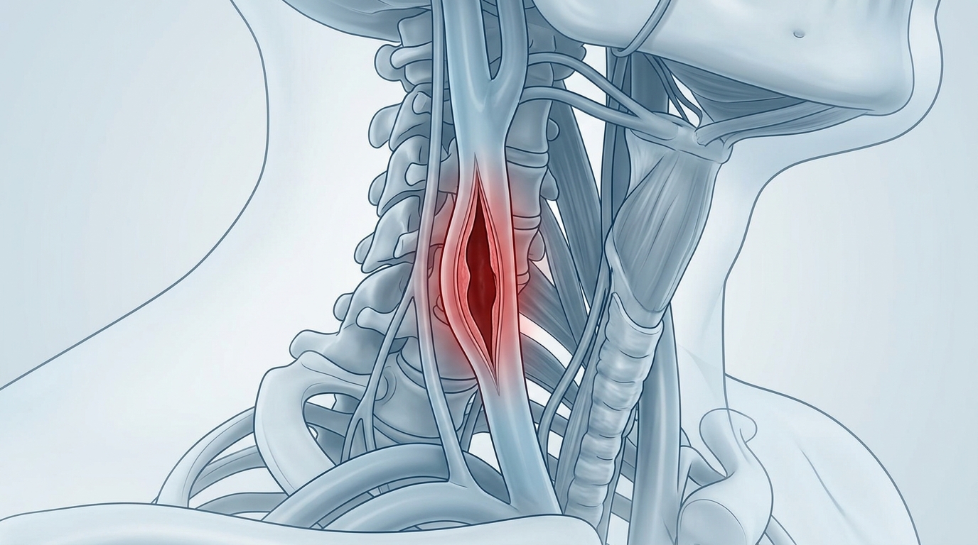
Carotid dissection refers to a tear in the wall of the carotid artery, the vessel that carries blood toward the brain. This lesion is associated with the appearance of Claude Bernard-Horner syndrome due to involvement of the neighboring sympathetic fibers. Clinically, this dissection typically presents with lateral neck pain and/or hemicranias, often accompanied by the distinctive ocular disturbance.
Its origin remains often multifactorial. One commonly references a preexisting arterial wall fragility, a minor previous trauma sometimes overlooked, a recent infection, or fluctuations in blood pressure related to episodic hypertension. It is precisely during the first two weeks following the onset of the painful Horner syndrome that the risk of ischemic stroke is greatest, justifying rapid management and close monitoring.
Recognizing the Warning in Time: What Patients and Doctors Should Remember
Diagnosis relies on urgent vascular imaging. Doppler ultrasound, CT angiography, and MR angiography are the reference tests that allow direct visualization of the arterial wall to confirm or rule out dissection. Once the diagnosis is made, management primarily relies on anticoagulant or antiplatelet therapy, with the clear objective of preventing an ischemic stroke from occurring.
In this care pathway, one actor plays a role often overlooked by the general public: the ophthalmologist. Frequently consulted first for a simple drooping eyelid, this specialist is actually on the front line for spotting warning signs and directing without delay toward appropriate cerebrovascular imaging. This clinical vigilance, discreet but decisive, illustrates how a symptom localized to the eye can reveal an issue that extends far beyond the realm of vision.
Behind a detail as seemingly banal as one eyelid being a little lower than the other can lie a true vascular emergency, especially in younger patients. Remembering that the neck pain associated with this ocular sign should always trigger alarm helps save precious time—the time that can separate effective care from an avoidable stroke. A good reason to never minimize what the body sometimes signals, even through the most discreet channels.