
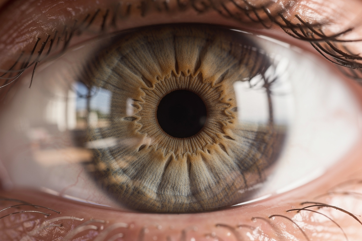
Before continuing, a small clarification is in order: this is not about an organ that can function without oxygen, but a tissue that has found a roundabout, nearly ingenious way to obtain it without going through the blood. We have long taught that blood irrigates absolutely everything, down to the smallest fold of skin or muscle. It’s also what we learn in the earliest biology classes: blood nourishes, oxygenates, repairs. Yet, tucked at the front of our eye, a transparent tissue defies this universal rule. Neither vein, nor artery, nor the slightest capillary feeds it, and yet it works perfectly, day after day, from birth to our final moments. This anatomical curiosity, far from a manufacturing flaw, is in fact a quiet triumph of evolution.
The cornea, this exception that defies the logic of the human body
The tissue in question is the cornea, that thin, curved membrane that covers the visible part of the eye and lets light pass before it reaches the lens and then the retina. Unlike all other tissues in the human body, the cornea is completely avascular, meaning it has no blood vessels, either large or small. This absence is not an anomaly or a developmental flaw; quite the opposite: it is essential to its primary function. If blood were to irrigate this tissue, red blood cells and the walls of the vessels would inevitably block the passage of light, making vision impossible.
We can thus grasp the importance of this peculiarity when we know that the cornea alone accounts for two-thirds of the eye’s refractive power, that is to say its ability to bend light rays to form a sharp image. A major optical role entrusted to a tissue that lives on the margins of the circulatory system tasked with nourishing every cell of our body.
How a living tissue survives without a single vein or artery
The question is almost vertiginous: how can living cells, which need nutrients and oxygen to function, survive without any direct blood supply? The answer rests in a single word: diffusion. The corneal cells draw what they need not from the blood, but from the surrounding media immediately around them. The edge of the tissue still benefits from a light support thanks to the limbal vascularization, this transitional zone located around the cornea at the junction with the white part of the eye. But for the rest, that is, almost the entire surface of the cornea, everything relies on passive exchanges.
The front surface of the tissue benefits from the tears, that thin film of tears that permanently covers the eye and acts as an interface with the surrounding air. The back surface, meanwhile, bathes in the aqueous humor, the clear liquid that fills the anterior chamber of the eye and that provides glucose and oxygen to the deeper layers. It is an extremely efficient system, but it remains fragile: without a backup blood source, the slightest disruption of these exchanges can soon put the tissue in trouble.
Oxygen from the air, a discreet fuel for a transparent organ
Here is perhaps the most surprising aspect of this story: a substantial portion of the oxygen that nourishes the cornea comes directly from the air we breathe, not from any internal circuit. The superficial layers of the tissue absorb this atmospheric oxygen through the tear film, somewhat like a plant captures sunlight to photosynthesize. The deeper layers, however, depend more on the oxygen dissolved in the aqueous humor. This double supply neatly illustrates the biological solution in place to compensate for the absence of vessels: the cornea oxygenates itself by diffusion from the air, not via the blood.
This mechanism, as elegant as it is, reveals its limits when an obstacle stands between the eye and the ambient air. This is precisely what happens with wearing contact lenses, so widely used today. By covering the cornea, the lens mechanically reduces access to atmospheric oxygen, which can lead, with prolonged use or improper fitting, to a slight swelling of the tissue or irritation at the limbus. There are indeed precise guidelines to assess the oxygen permeability of different lens materials, precisely to limit such discomfort.
What this anatomical oddity reveals about the fragility of our eye
This absence of vessels, clever as it is, has a downside. In the face of a severe assault, such as a deep infection, a burn, or significant trauma, the cornea cannot rely on a rapid influx of blood to compensate for a sudden oxygen deficit. It is then exposed to a risk of hypoxia, a deprivation of oxygen that can permanently undermine the tissue if nothing is done to address it.
In some cases, the body even attempts to respond by developing corneal neovascularization, that is, the growth of new tiny vessels that gradually invade the tissue. A reaction that starts with good intentions—to better feed the distressed area—but backfires on the eye: these capillaries, by settling where they normally do not belong, compromise the very transparency sought and can alter vision in a lasting way. An almost poetic paradox, where the solution meant to save the tissue ends up threatening its most essential function.
Ultimately, the cornea reminds us that our body does not always operate according to a single universal rule. This small transparent disk, invisible to our daily concerns, stands thanks to a delicate balance between air, tears, and a discreet ocular fluid. A silent, impressive feat that deserves a moment of thought the next time we blink, or remove our contacts after a long day.