
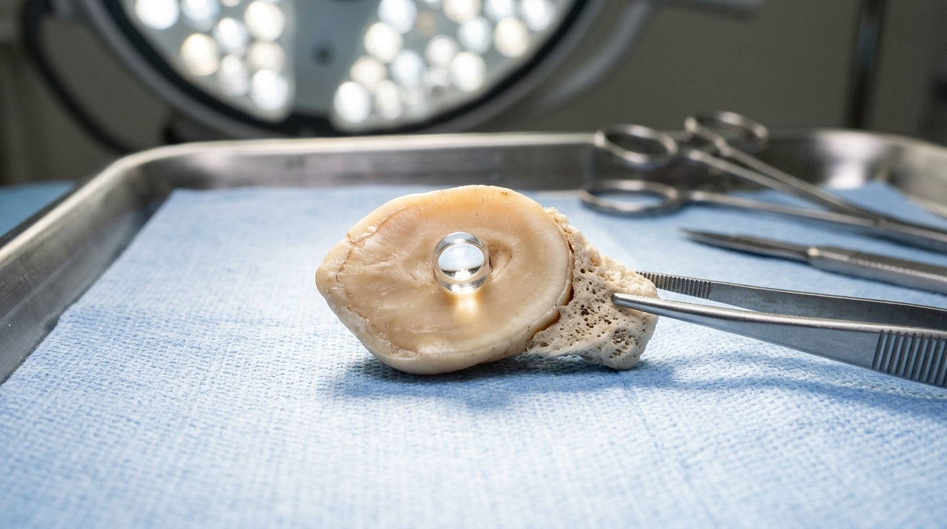
Implanting a tooth into the eye of a blind patient to restore sight: it sounds like something straight out of a science fiction novel. Yet this technique has existed since the 1960s, and today it continues to save sight where modern medicine falls short.
- The osteo-odonto-keratoprosthesis (OOKP), developed by Benedetto Strampelli in the 1960s, involves implanting a canine tooth and its supporting bone into the eye to secure an optical cylinder, because dentin helps prevent implant rejection.
- In February 2025, Vancouver’s Mount Saint Joseph Hospital performed the two-stage procedure in three formerly blind patients, including Brent Chapman, who regained vision to various extents.
- A 2005 study showed an 85% implant survival rate after 18 years, a figure later corroborated by a 2022 study reporting 94% anatomical survival up to 30 years of follow-up.
- A Tooth to See: The Bold Idea That Defied Medicine
- Canine, Jawbone and Optical Cylinder: The Anatomy of a Surgical Miracle
- 85% Success After 18 Years: The Figure That Validated the Unthinkable
Tooth to See: The Bold Idea That Defied Medicine
Imagine losing your sight not due to a retinal problem or optic nerve issue, but because the surface of your eye—the cornea—has become entirely opaque. In such a case, a standard corneal graft would not work, since the recipient tissue can no longer accommodate a transparent cornea. This is precisely the barrier some patients faced more than sixty years ago.
That is where Benedetto Strampelli, an Italian surgeon, steps in with a solution as audacious as it is ingenious. His reasoning is simple on paper: since the body rejects almost all foreign materials placed inside the eye, why not use tissue that the body already recognizes as its own? His answer comes in one word: a tooth.
The technique carries a harsh name, the osteo-odonto-keratoprosthesis, often abbreviated as OOKP. Behind this term lies a surgical feat: removing a canine tooth from the patient along with a portion of its supporting bone to serve as the natural scaffold for an artificial optical lens. The principle has endured through the decades with little real competition.
Evidence that this idea remains relevant was seen recently in Canada. In February 2025, Mount Saint Joseph Hospital in Vancouver carried out the first stage of an OOKP never before performed in the country, on three patients who had been blind for many years.
Canine, Jawbone and Optical Cylinder: The Anatomy of a Surgical Miracle
Practically speaking, how can a tooth help an eye to see? It hinges on a material you might not suspect could be so valuable in ophthalmology: dentin. According to Dr. Greg Moloney, who led the Vancouver operation alongside maxillofacial surgeon Shannon Webber, it is precisely this dental tissue that prevents the body from rejecting the plastic optical implant.
The procedure unfolds in two clearly separated stages, spaced by several months. First, surgeons harvest a canine tooth along with a portion of its supporting bone, then shape and drill it to accommodate a small optical cylinder. This assembly is then buried in the patient’s cheek. This step allows the structure to vascularize, i.e., develop its own blood supply, before it is transplanted.
Once vascularization is established, the second stage begins. The tooth-bone-lens block is carefully extracted from the cheek and implanted into the eye, replacing the cornea that has become opaque. The tooth root and jawbone form a stable biological frame that surrounds the fixed optical cylinder, giving the eye a durable anchor that the body accepts over the long term.
Brent Chapman’s case illustrates the value of this approach for the most desperate situations. This 33-year-old Canadian, who became blind in adolescence after a severe reaction to an anti-inflammatory drug, had undergone around fifty unsuccessful procedures before receiving the OOKP. In July 2025, the hospital announced that he and the other two operated patients could see again, albeit to varying degrees.
85% Success After 18 Years: The Figure That Validated the Unthinkable
A technique so spectacular would deserve to be dismissed if it rested on only a handful of cases. Yet decades of data tell a different story. A 2005 study by Falcinelli in the Archives of Ophthalmology followed 181 cases over the long term, a sample large enough to draw solid conclusions.
The result speaks for itself: 85% of implants remained in place 18 years after the initial operation. For a prosthesis implanted in an environment as demanding as the human eye, that long-term survival rate is almost astonishing for its consistency. A more recent retrospective study, published in 2022 on 82 eyes treated with Strampelli’s original technique, confirms this trend with a 94% anatomical survival rate up to 30 years of follow-up.
France has not been a stranger to this surgical venture. As early as 2014, the CHU of Marseille, through the ophthalmology department at La Timone led by Prof. Louis Hoffart and the maxillofacial department at Nord hospital, operated on three patients with bilateral corneal blindness using the same method.
On the funding side, the Canadian program came to life thanks to a donation from the St. Paul’s Hospital Foundation, which gathered $430,000 for its first three years. A substantial investment, but one that seems justified given the outcomes in patients once deemed incurable.
Sixty years after its invention by Benedetto Strampelli, the osteo-odonto-keratoprosthesis continues to prove its relevance, away from the limelight yet with an effectiveness that commands respect. It reminds us of a simple truth: sometimes the most avant-garde solutions take the most unexpected routes, turning a tooth into a window onto the world.
👉 A small gesture for Space Group? Follow us on Google, add us to your bookmarks and find us on Google News so you won’t miss any discovery.