
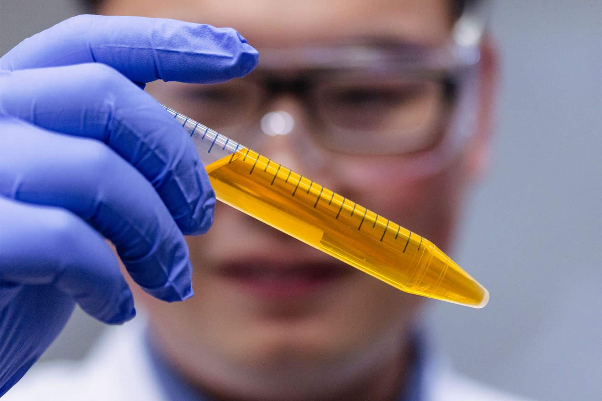
Who could have imagined that such a common ingredient might revolutionize biology? A team of researchers has recently discovered that a common food dye can render mouse skin translucent. Yet, it remains unclear whether this approach would work in humans. This discovery, at any rate, opens up new possibilities for medical imaging and biological research.
The dance of light in biological tissues
The innovation behind this method rests on a deep understanding of how light interacts with biological matter. Our tissues are made up of a multitude of molecules, each possessing a specific refractive index. This index determines how light propagates through the material: it can be absorbed, reflected, or refracted (that is, bent from its path).
When light traverses a biological tissue, it encounters interfaces between different substances (water, proteins, lipids, etc.), each with its own refractive index. At every interface, part of the light is scattered in all directions, which makes the tissue opaque to the eye. It’s a bit as if the light is perturbed along its journey, preventing a clear view of what lies beneath.
The role of the food dye: a delicate balancing act
The researchers conceived using this property of light to render tissues transparent, or rather translucent. By applying a specific food coloring, they modified the tissues’ refractive index, thereby smoothing the propagation of light through them.
The Tartrazine, a yellow dye widely used in the food industry, was chosen for this experiment. By penetrating the tissues, this dye binds to proteins and other molecules present, thereby altering their optical properties. Thanks to this modification, light is less scattered and can pass through the tissues more uniformly, making the internal structures visible.
A window into the living organism
This technique offers a new perspective on studying living organisms. By making the skin transparent, it indeed enables real-time observation of the biological processes taking place inside the body. The researchers have thus been able to visualize blood flow in the smallest vessels, which is essential for studying circulation and cardiovascular diseases. Organs such as the liver, kidneys, or intestines have also become visible, allowing the study of their structure and function. Finally, muscle contractions could be observed in detail, which could revolutionize the study of muscular physiology.
Promising medical applications
This discovery opens the door to numerous medical applications. Imagine being able to visualize veins with greater precision to facilitate blood sampling or to detect cancerous tumors earlier by directly observing the tissues involved. This technique could also revolutionize basic research by enabling real-time study of disease development and the effectiveness of new treatments.
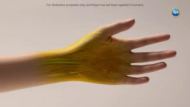
Although this technique has been successfully tested on mice, many questions remain before it can be applied to humans. Human skin is thicker than that of rodents, which could make dye absorption more challenging. Moreover, it will be necessary to ensure that this method is entirely safe and free from long-term side effects.
Nevertheless, researchers are optimistic about the prospects this discovery offers. They believe this technique could become an essential tool for clinicians and biologists, opening new avenues for diagnosing and treating diseases.