
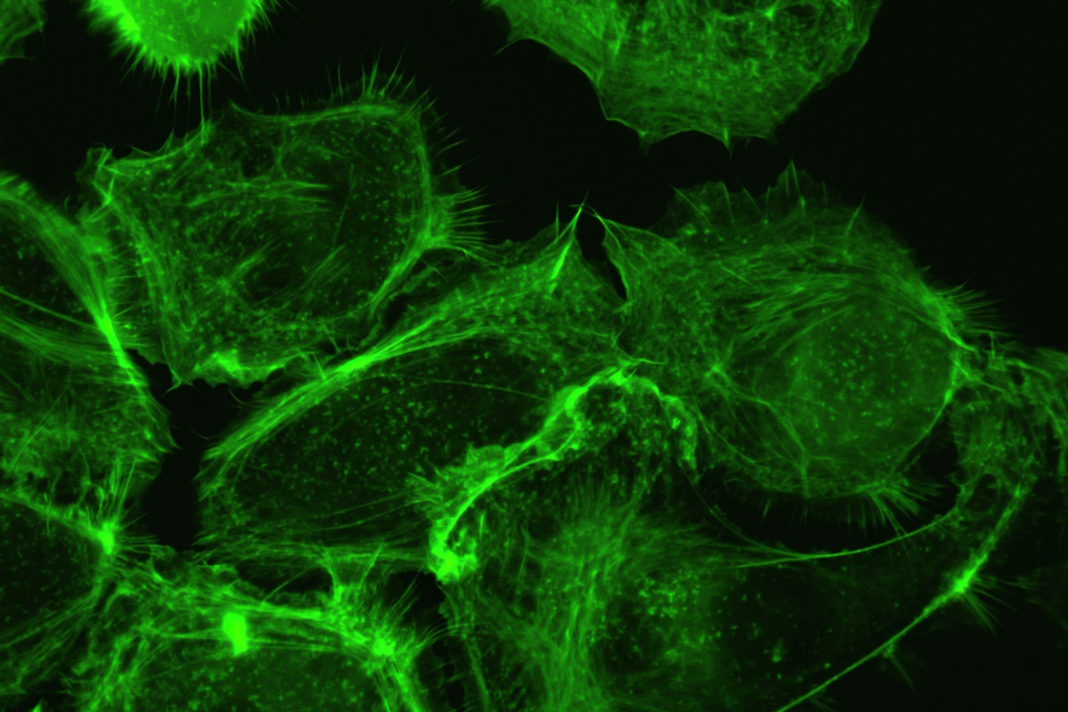
The biological cell is the fundamental unit of all living organisms. For many decades, however, observations with conventional light microscopy were akin to trying to watch a bustling city at night without street lighting: silhouettes and outlines could be discerned, but the dynamic processes happening inside remained hidden in the dark. It was only with targeted use of fluorescence that biomedical research underwent a radical transformation. Today, this technology enables real-time, three-dimensional tracking of molecular processes with fascinating precision.
The Physical Principle: Light on Demand
To understand the phenomenon of fluorescence, it is useful to examine the physical principles of light absorption and emission. Fluorescence refers to the property of certain substances (called fluorophores) to absorb light at a specific wavelength and to re-emit almost instantaneously light at another wavelength, with lower energy.
The process unfolds in three essential phases:
- Excitation: a fluorophore absorbs energy in the form of light (for example, blue or ultraviolet light). This causes the electrons of the molecule to be excited to a higher energy level.
- Energy loss: A small portion of the absorbed energy is dissipated as heat through molecular vibrations.
- Emission: When the electrons fall back to their ground state, the remaining energy is emitted as light. Because part of the energy has been lost, the emitted light has lower energy and therefore a longer wavelength (for instance, green or red light).
This difference between the wavelength of the incident excitation light and that of the emitted light is called the Stokes shift. It is the physical key to microscopy: optical filters can completely block the excitation light, so that only the signal emitted by the samples is detected. The result is an image of exceptional sharpness and high contrast on a dark background.
The Historical Turning Point: a Jellyfish Protein That Changed the World
Although fluorescent dyes were known as far back as the 19th century, the true breakthrough in the life sciences occurred in the 1960s with the discovery of green fluorescent protein (GFP). This protein was isolated from the jellyfish Aequorea victoria.
The scientific breakthrough followed in the 1990s: researchers discovered that the GFP genetic code could be fused to the genes of other organisms. When a cell produces a specific target protein, it also produces the fluorescent GFP.
The green glow then acts as a molecular beacon. For the first time, it was possible:
- To determine the exact location of proteins in living cells.
- To observe the transport of molecules across cell membranes.
- To monitor in real time cell division, differentiation, and movement.
For this discovery and its subsequent developments, the 2008 Nobel Prize in Chemistry was awarded to Osamu Shimomura, Martin Chalfie and Roger Tsien. Today, laboratories around the world possess a full range of genetically engineered proteins that fluoresce in every color of the rainbow.
Medical Application Domains: From the Laboratory Bench to the Operating Room
Fluorescence analysis has become an indispensable tool in nearly all areas of modern medicine.
Cancer Research and Tumor Diagnostics
In oncology, fluorescence enables a more precise characterization of early stages of tumor diseases. Cancer cells often display altered surface proteins. If these are targetedly labeled using fluorescent antibodies, residual tumor cells can be identified even when they form only tiny clusters.
This method is also used in surgery (fluorescence-guided surgery): before an intervention, patients receive a well-tolerated fluorescent dye that accumulates mainly in tumor tissue. Under a specific illumination, the surgical team can see exactly where diseased tissue ends and where healthy tissue of the organ begins.
Neuroscience and Brain Mapping
The human brain comprises billions of interconnected neurons. With fluorescent calcium sensors, it is possible to visualize the electrical activity of nerve cells. As soon as a neuron fires a signal, calcium ions flow in, causing a change in fluorescence intensity. This allows the study of complex neural networks and learning processes in living tissue.
Infection Biology and Virology
In the study of pathogens such as viruses or bacteria, fluorescence helps to understand how these agents bind to host cells and penetrate them. Using multi-marker techniques, it is possible to label simultaneously and distinctly the virus, the cell membrane, and the cellular transport pathways.
High-Throughput Analytics: Large-Scale Fluorescence Measurement
While microscopy provides a visual snapshot of individual cells, pharmaceutical research requires the analysis of thousands of samples in a short time. In the search for new drugs, enormous libraries of substances must be tested to determine whether they inhibit or stimulate biochemical reactions.
For this purpose, microplates are used—a plastic plate with dozens, or even thousands, of small wells. To quickly and quantitatively detect fluorescence signals across all these samples, specialized measuring devices are employed, such as a fluorescence microplate reader. These instruments handle the light excitation and signal detection in a highly automated manner, allowing researchers to obtain precise data within minutes on enzymatic activities, protein interactions, or cell viability.
Evolution: Pushing Beyond the Limits of Physics
Classical optical microscopy long confronted a physical limit: due to the diffraction of light, it seemed impossible to distinguish structures smaller than about 200 nanometers (the Abbe diffraction limit).
Thanks to innovative fluorescence techniques, this barrier has nonetheless been breached:
- Super-resolution microscopy (for example, STED, STORM): by selectively activating and deactivating neighboring fluorescent molecules, it is possible to achieve resolutions on the order of a few nanometers. This makes individual protein complexes visible.
- Optogenetics: by combining fluorescence labeling techniques with photosensitive proteins, cellular processes are no longer just observed, but can be controlled in a targeted manner using light.
Fluorescence is much more than a mere aesthetic optical phenomenon of nature. Through the synthesis of physics, chemistry, and molecular biology, it has become one of the most powerful tools in medical diagnostics and disease research.
Today, analyses can also be made even more powerful thanks to artificial intelligence. It greatly increases the speed at which data can be analyzed and compared. The PANDA technology shows how AI helps extract information from high-resolution images and thus contributes to tumor detection. The ability to make the invisible visible is daily laying the foundations for new therapies, more targeted treatments, and a deeper understanding of the biological processes of life.