
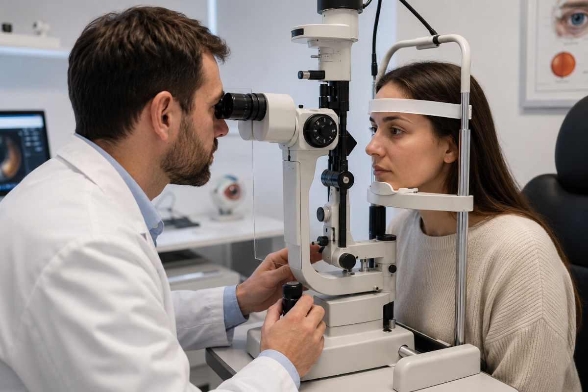
Despite the growth of OCT, retinal imaging, and digital imaging systems, the slit lamp retains a central role in ophthalmic examination. This biomicroscope makes it possible to observe directly, at various magnifications and lighting, a multitude of eye structures. This versatility helps explain why the instrument remains part of the daily practice of ophthalmologists and, depending on their expertise and practice setting, of other vision professionals.
A Direct Examination That Is Particularly Rich
The slit lamp combines a binocular microscope with a light source whose beam can be adjusted in height, width, and orientation. The practitioner thus gains a magnified, three-dimensional view of the observed structures. The examination primarily targets the eyelids, the conjunctiva, the cornea, the anterior chamber, the iris, and the lens. With the appropriate lenses, the exploration can also extend to the vitreous, the retina, or the optic nerve. This ability to switch rapidly from one structure to another is one of the main clinical advantages of the device. The Merck Manual specifically notes its usefulness for detecting corneal abnormalities, assessing the depth of the anterior chamber, and localizing opacities in the lens.
Modulable lighting provides information that a simple eye photograph cannot supply. Depending on the angle and thickness of the beam, the professional can examine different corneal layers or search for inflammatory signs in the anterior chamber. The addition of fluorescein also helps to better visualize certain surface lesions. This dynamic aspect is highly valuable in practice: the practitioner immediately adapts the observation to what is found and can probe a suspicious area more deeply without completely changing the examination apparatus.
Indications at the Heart of Consultations
The slit lamp contributes to the assessment of a wide range of common symptoms: red eye, pain, foreign-body sensation, reduced vision, or discomfort related to contact lens wear. It can reveal involvement of the conjunctiva or cornea, intraocular inflammation, or lens opacification compatible with cataract. It also plays a role in the follow-up of chronic diseases, because repeated clinical examination allows monitoring the appearance, persistence, or evolution of certain signs.
This role remains clearly visible in contemporary guidelines. The American Academy of Ophthalmology incorporates the biomicroscopic slit-lamp examination into the comprehensive medical evaluation of the adult eye, alongside pupil examination, intraocular pressure measurement, and fundus examination. The device thus does not replace OCT, retinal photography, or other supplementary tests. It provides a distinct clinical piece of information, directly accessible during the consultation, and helps determine which additional tests are truly necessary.
A Device That Continues to Evolve
The age of the underlying principle does not mean the instrument has stood still. Modern models can incorporate several magnifications, different filters, and digital imaging solutions designed to keep a record of the examination. The ability to record images is particularly valuable for documenting a lesion, comparing its evolution across visits, or facilitating exchanges between professionals. Ergonomics and optical quality are also important, since precise examination requires stable adjustment and comfortable observation, even with repeated consultations.
For opticians, ophthalmologists, or orthoptists seeking to equip an examination space within their scope of practice, choosing a slit lamp depends less on an accumulation of features than on the intended uses. Visionix offers equipment aimed at vision professionals, with various magnification options and, depending on the model, digital imaging capabilities. It remains useful to assess optical quality, the precision of adjustments, patient and operator comfort, space requirements, and documentation possibilities. The budget should also cover the necessary accessories, maintenance, and the practical organization of the examination station.
A Clinically Central Tool
The slit lamp remains essential because it meets a simple need: to directly observe the eye with precision and from multiple angles. Imaging technologies today enrich diagnosis, but they do not render this direct observation obsolete. In everyday practice, its versatility, quick usability, and the richness of information gathered explain its enduring value.